
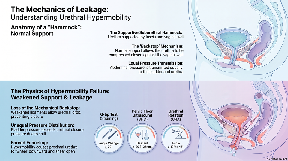


What is urethral hypermobility, and how does it physically cause stress urinary incontinence?

What is urethral hypermobility, and how does it physically cause stress uri... | WHC Clinical FAQ

How do clinicians differentiate stress urinary incontinence from overflow incontinence caused by...
Urinary assessment
Mechanism-led
Safety first
Women’s Health Clinic FAQ
What is urethral hypermobility, and how does it physically cause stress urinary incontinence?
Stress urinary incontinence can feel straightforward, but the clinical cause may involve urethral support, sphincter closure, bladder behaviour, prolapse, tissue quality or a mimic that needs a different pathway.
Direct answer
Urethral hypermobility means the urethra and bladder neck move too far when pressure rises during coughing, laughing, lifting or exercise. The pelvic-floor and fascial support system normally gives the urethra a stable backing so it can close under pressure. When that support is stretched or weakened, pressure is transmitted unevenly and urine may leak even though the bladder muscle is not contracting.
The safest answer comes from matching the symptom pattern with examination and appropriate tests, rather than assuming that every leak has the same cause or needs the same treatment.
Educational only. This page is educational and does not replace individual assessment. Results vary. Not a cure.

Assessment-led continence care
At a glance
These are the main clinical points behind this question.
At a glance
Clinical summary
Core mechanism
Stress leakage often reflects support, closure or both.
Structures involved
Urethra, bladder neck, sphincter, mucosa, fascia and pelvic floor.
Assessment value
Mechanism helps guide conservative, medical or specialist options.
Key limit
Severity alone does not prove the underlying mechanism.
Important safety note
Seek medical advice promptly for blood in urine, recurrent infection symptoms, urinary retention, new neurological symptoms, severe pelvic pain, a pelvic mass or postmenopausal bleeding.
Examination
Testing
Mechanism
Review
Detailed answer
Detailed answer
Continence depends on a stable urethral support system and an internal seal that can resist sudden pressure rises.
Clinical context
Hypermobility describes movement and support failure; sphincter deficiency describes reduced closure strength.
Bladder
Pelvic floor
Prolapse
What it means
Continence depends on a stable urethral support system and an internal seal that can resist sudden pressure rises.
Why it happens
Hypermobility describes movement and support failure; sphincter deficiency describes reduced closure strength.
Where testing helps
Mucosal coaptation, vascular cushioning and tissue quality can influence how well the urethra seals.
What it cannot prove
A careful assessment prevents all stress leakage being reduced to a single explanation such as weak muscles.
What this means in practice
A continence plan should be based on the likely mechanism, symptom burden and safety checks, not on a single label.
If symptoms are mixed, new, painful or difficult to explain, a more detailed assessment is usually safer than rushing into treatment.
Patient safety
Why this distinction matters
Different causes of urinary leakage need different priorities, and the wrong assumption can lead to poor expectations or the wrong care pathway.
It clarifies the cause
Stress, urge, overflow, prolapse-related and urethral causes can overlap but are managed differently.
It protects safety
Blood in urine, retention, recurrent infection symptoms, severe pain or neurological symptoms should not be treated as routine SUI.
It shapes treatment
Pelvic-floor therapy, bladder treatment, pessary support, medication review or specialist referral may be considered depending on findings.
It sets expectations
A clear diagnosis helps explain what improvement is realistic and when reassessment is needed.
A mechanism-led approach
Good continence care separates the trigger, the anatomy, the bladder response and the patient's quality-of-life burden.
This makes the final discussion more useful than a simple yes-or-no answer.
Considerations
What to consider
The right next step depends on symptoms, examination, bladder emptying, prolapse findings, tissue health and how much the leakage affects daily life.
Consultation priorities
The clinician asks about triggers, childbirth history, pelvic surgery, menopause, coughing, exercise and symptom burden.
Emptying
Prolapse
Goals
History
The clinician asks about triggers, childbirth history, pelvic surgery, menopause, coughing, exercise and symptom burden.
Examination or tests
Examination may assess prolapse, urethral mobility, pelvic-floor function and visible stress leakage.
Interpretation
Additional tests are considered when mechanism is unclear or treatment planning would change.
Next steps
The plan may include pelvic-floor therapy, pessary support, medical review or referral where appropriate.
Practical expectations
Some women need only conservative assessment and pelvic-floor support; others need bladder, prolapse, urology or urogynaecology review.
Costs, access and treatment details should be confirmed directly with the clinic before booking.
Common concerns and myths
Common misconceptions
These points help prevent over-simple explanations of stress urinary incontinence.
Myth: SUI is only weak pelvic-floor muscle
Reality: support, sphincter closure, mucosal seal and tissue quality can all contribute.
Myth: hypermobility and sphincter deficiency are the same
Reality: they can overlap, but they describe different parts of continence.
Myth: severe leakage proves the cause
Reality: severity describes burden, not the whole mechanism.
Evidence and uncertainty
Clinical findings can be useful without being perfect; the safest wording is clear about limits.
Personalised planning
The most suitable pathway depends on mechanism, symptom burden, health history and the patient's priorities.
Safety checklist
Safety checklist
Use these questions to decide whether continence symptoms need routine review, more detailed assessment or urgent advice.
Is the trigger clear?
Stress leakage is usually linked to cough, laugh, lifting, running or sudden pressure.
Is emptying normal?
Weak stream, incomplete emptying or constant dribbling may suggest retention or overflow rather than simple SUI.
Are red flags absent?
Seek medical advice promptly for blood in urine, recurrent infection symptoms, urinary retention, new neurological symptoms, severe pelvic pain, a pelvic mass or postmenopausal bleeding.
Is the plan reviewed?
If symptoms change, persist or do not fit the expected pattern, reassessment is more useful than repeating the same approach.
Reassuring signs
Symptoms are easier to interpret when triggers are consistent, urine testing is clear, emptying feels normal and there are no new red flags.
No red flags
Review plan
Reasons to pause
Seek medical advice promptly for blood in urine, recurrent infection symptoms, urinary retention, new neurological symptoms, severe pelvic pain, a pelvic mass or postmenopausal bleeding.
Retention
Pain
When to escalate
When to seek medical help
Some urinary symptoms should be checked before they are labelled as stress incontinence.
Use NHS 111 online
Blood in urine
Visible blood or unexplained blood on urine testing needs medical assessment.
Retention or overflow signs
A weak stream, inability to empty, increasing bladder discomfort or constant dribbling should be reviewed.
Infection or severe pain
Fever, flank pain, burning with systemic symptoms, severe pelvic pain or feeling unwell needs prompt advice.
Emergency symptoms
Call 999 in a life-threatening emergency, including collapse, chest pain or breathing difficulty.
Use NHS 111 for urgent advice or call 999 in a life-threatening emergency. This page is educational and does not replace individual medical assessment.
Regulatory resources
Authoritative resources
These resources support assessment-led, evidence-aware continence information.
NICE NG123: urinary incontinence and pelvic organ prolapse
NICE anchors UK standards for urinary incontinence assessment and management.
NHS: urinary incontinence
NHS patient information supports clear descriptions of stress, urge and overflow leakage.
BAUS: stress urinary incontinence
BAUS gives urology context for SUI assessment and treatment discussions.
Next step
Book a continence consultation
A consultation may help clarify the likely cause of leakage, whether tests are needed and which treatment or referral pathway is most appropriate.
▶ View Research Sources (12 Sources)
These 12 source names are selected from 60 curated sources. Additional reviewed material included professional society guidance, peer-reviewed clinical papers, clinical trial records; duplicate and low-relevance records were removed before display.
Educational only. This information is for education only and is not a substitute for professional medical advice, diagnosis or treatment. Results vary. Not a cure.